
FileNeuron1.jpg Simple English Wikipedia, the free encyclopedia
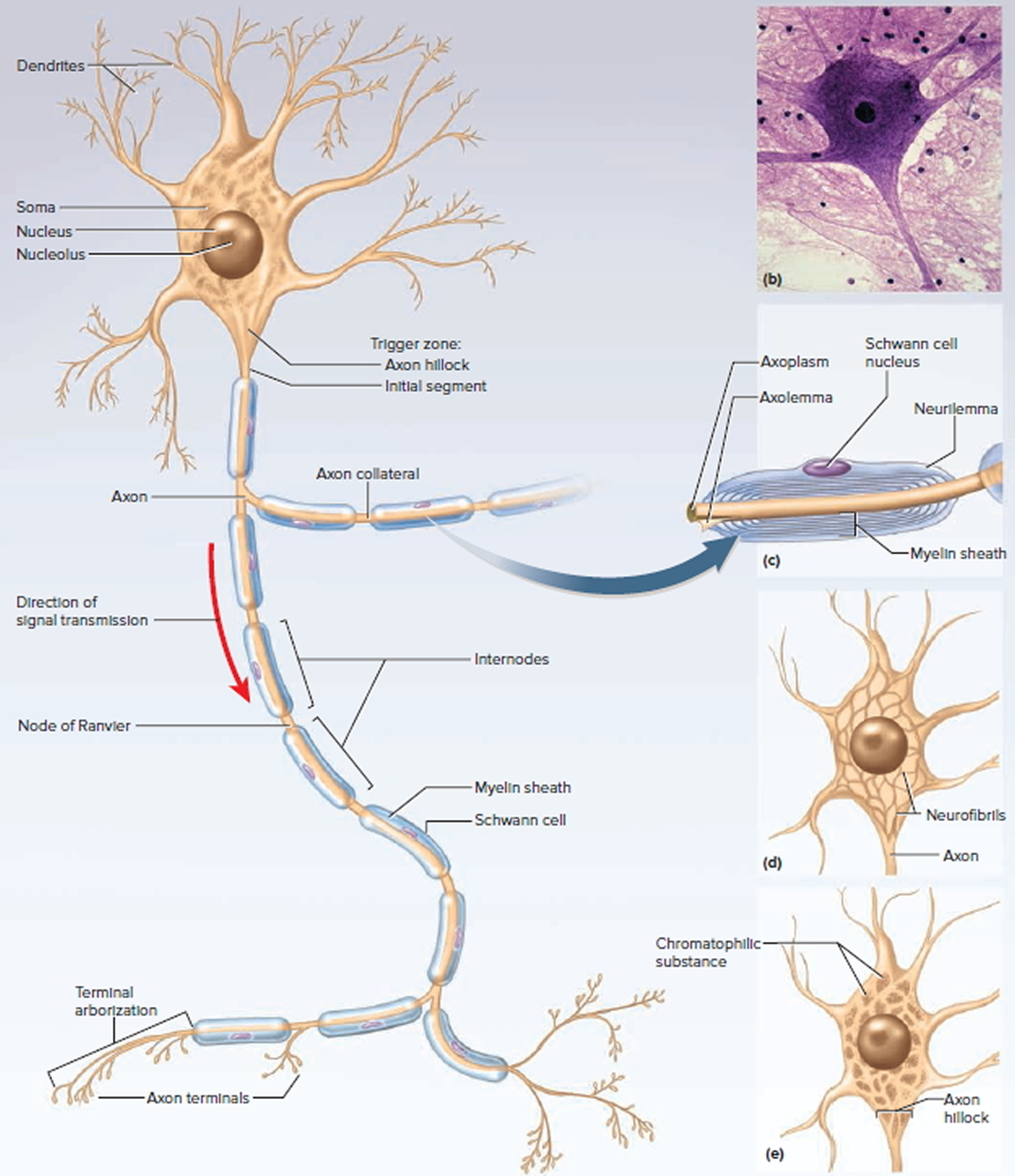
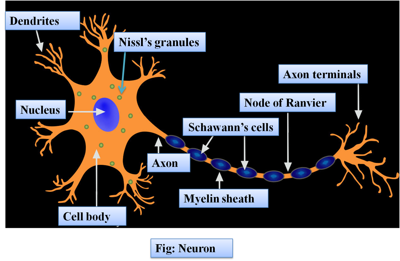
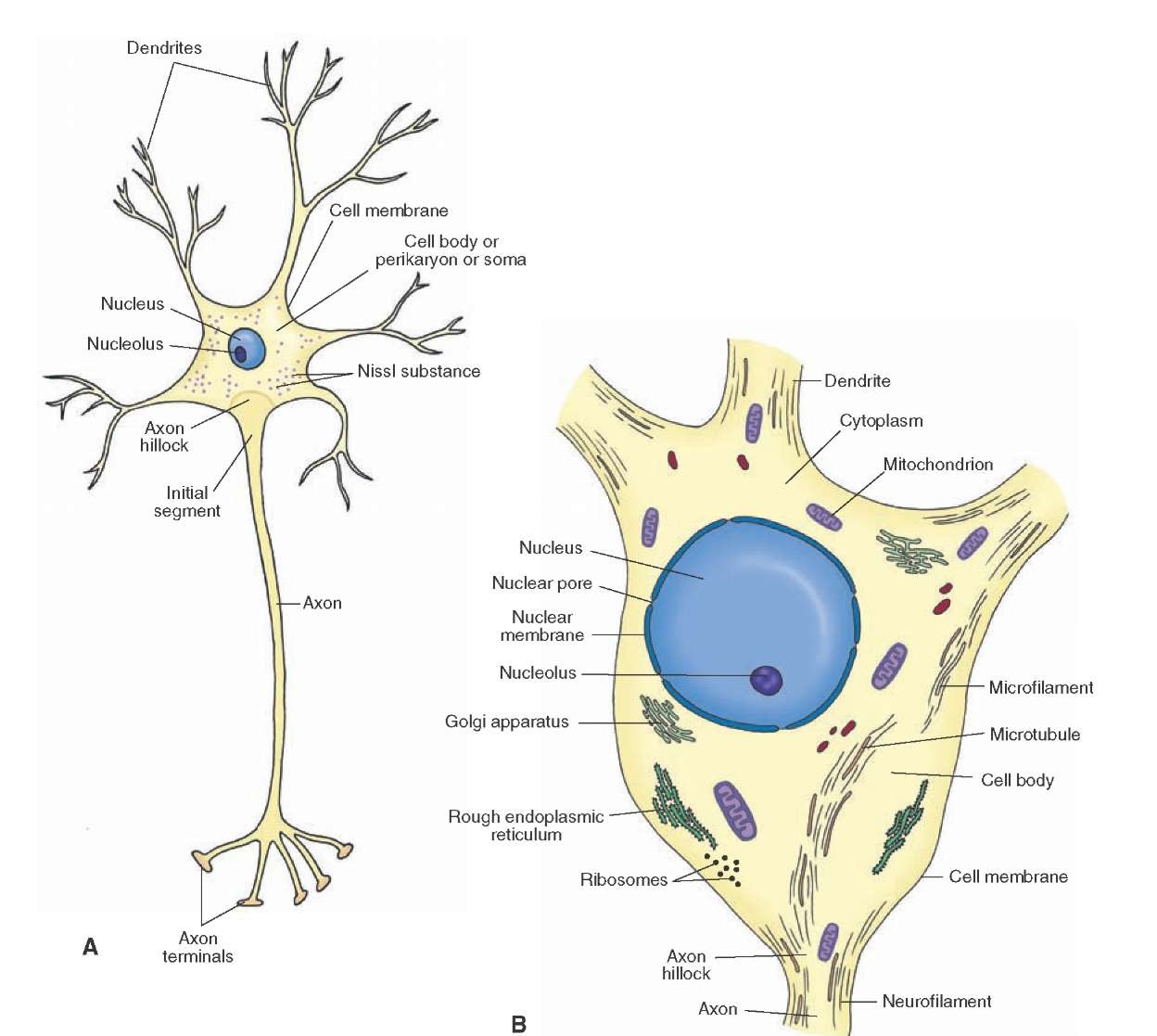
Diagram Of Neuron. A neuron is a specialized cell, primarily involved in transmitting information through electrical and chemical signals. They are found in the brain, spinal cord and the peripheral nerves. A neuron is also known as the nerve cell. The structure of a neuron varies with their shape and size and it mainly depends upon their.
Neurons The crazy wires in our body. Doc Jana
What is the structure labeled 13? (orange dots throughout the cell) nissil substance. What is the circled structure? motor end plate. Study with Quizlet and memorize flashcards containing terms like What is the structure labeled 1?, What is the structure labeled 2?, What is the structure labeled 3? and more.
SCB209 Lab1 Natural Sciences Open Educational Resources
Like the heart, lungs, and stomach, the nervous system is made up of specialized cells. These include nerve cells (or neurons) and glial cells (or glia ). Neurons are the basic functional units of the nervous system, and they generate electrical signals called action potentials, which allow them to quickly transmit information over long distances.
Nerve Cell Diagram Labeled ClipArt Best
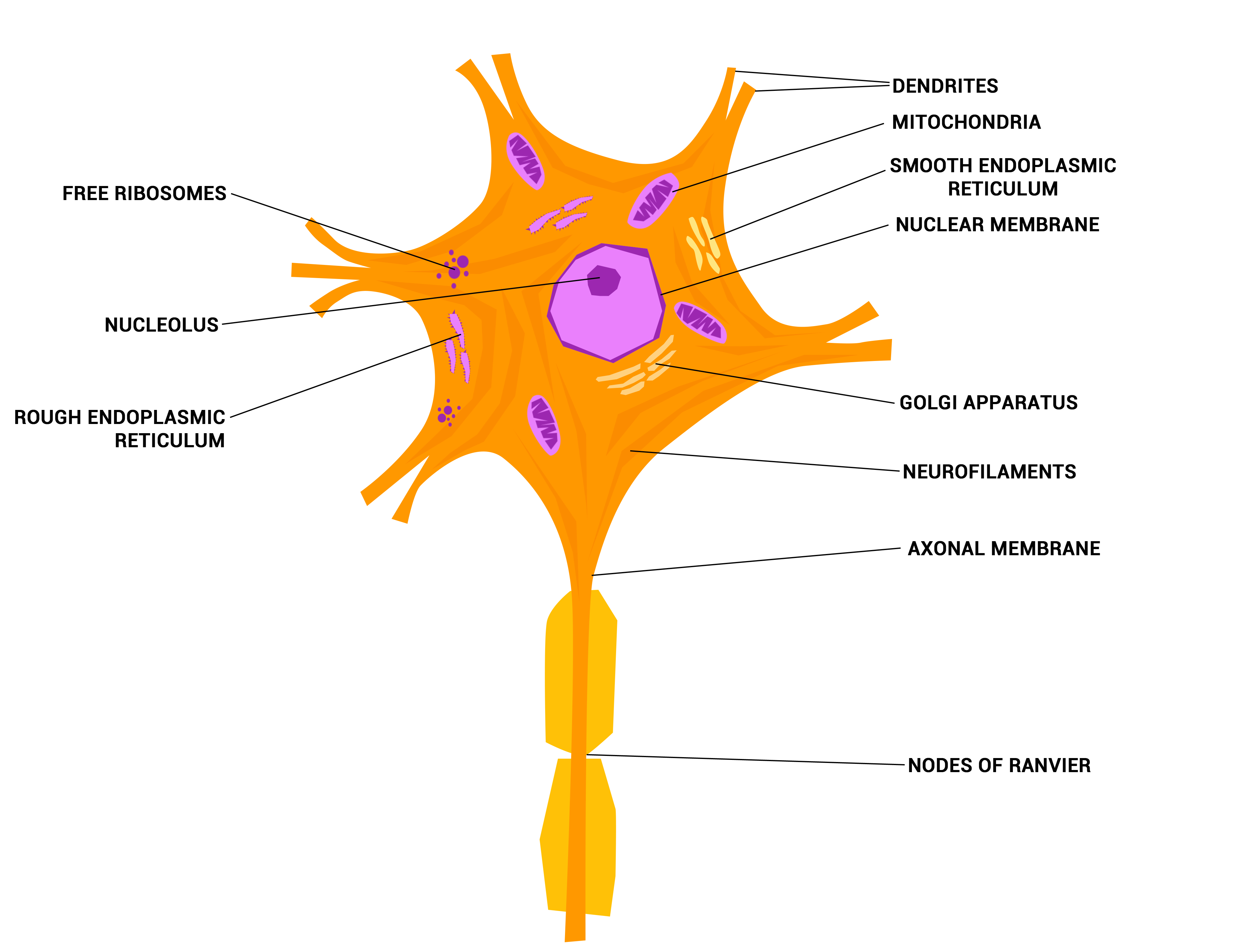
While they have the common features of a typical cell, they are structurally and functionally unique from other cells in many ways. All neurons have three main parts: 1) dendrites , 2) cell body or soma, and 3) axons. Besides the three major parts, there is the presence of axon terminal and synapse at the end of the neuron.

neuron model labeled Diagram Quizlet
Figure 12.2.2 - Parts of a Multipolar Neuron: The major parts of the neuron are labeled on a multipolar neuron from the CNS. External Website. Visit this site (link not working as of 10/20/2021) to learn about how nervous tissue is composed of neurons and glial cells. Neurons are dynamic cells with the ability to make a vast number of.

Neuron Model Labeled
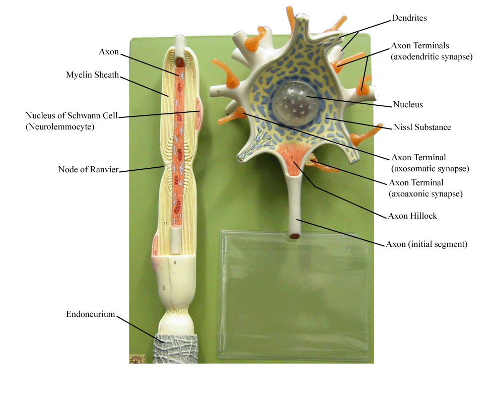
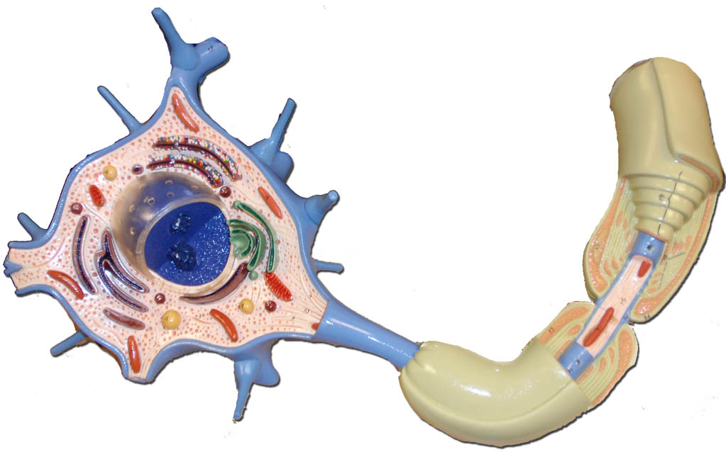
For pictures of this model with answer keys to help you study, visit:http://www.humanbodyhelp.com/brain-and-spinal-cord/http://www.humanbodyhelp.com/neuron-m.

Nervous and Spinal Cord Models
How to label a neuron using the best practices. There are a few different ways to label a neuron, but using a neuron chart is the most accurate and efficient way. A neuron chart is a graphical representation of brain cells, making labeling neurons easy and precise. A neuron chart can be made with paper or online and customized to fit your needs.
A diagram of a neuron and its functions. a study in chartreuse
Neuron Models. Click on a photo for a larger view of the model. Click on Label for the labeled model. Back to Nervous System. Neuron. Cell Body & Dendrites. Axon.
Nervous System Central and Peripheral Nervous System Function
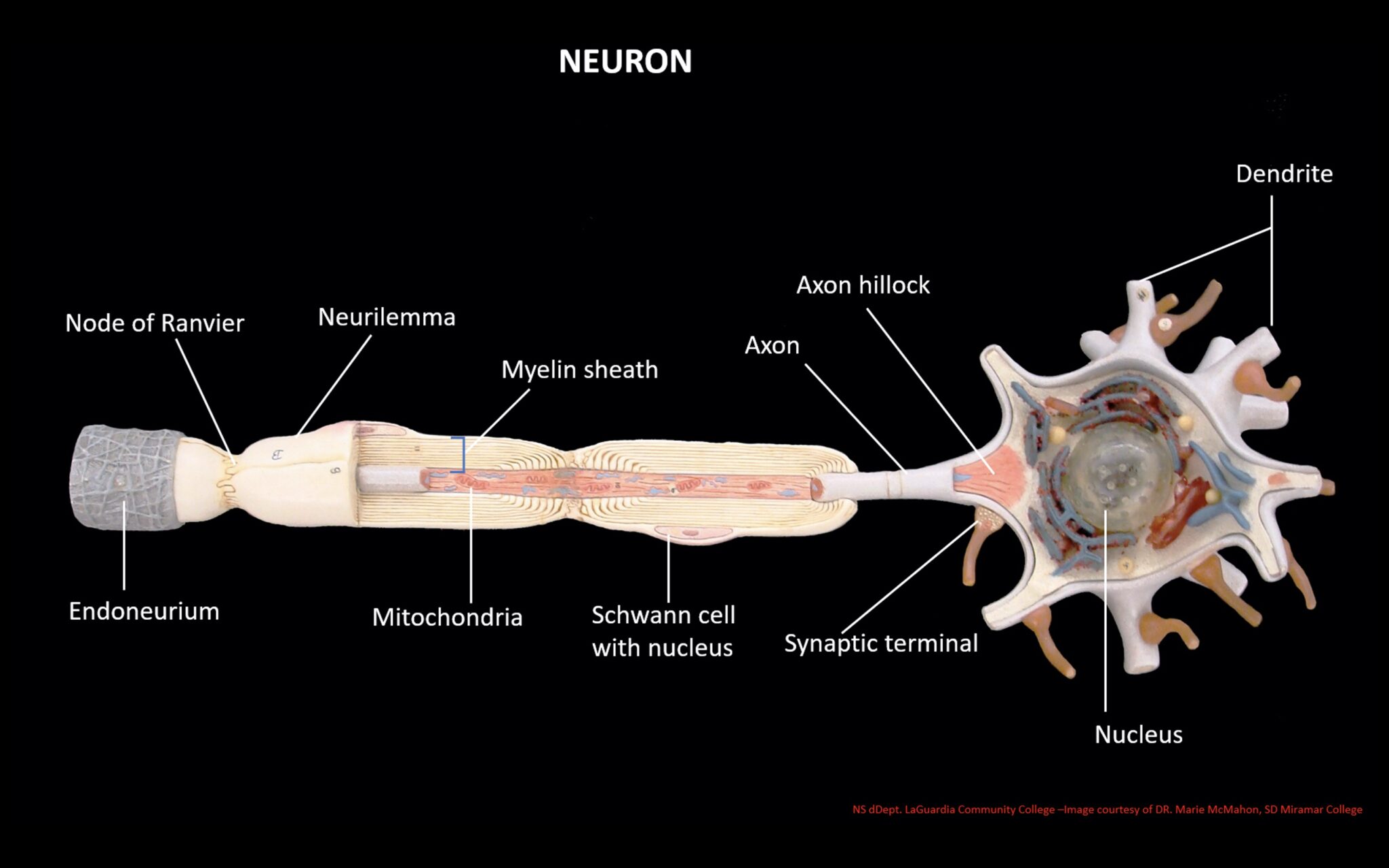
The structure of a motor neuron is characterized by three components: the soma, the axon, and the dendrites. Motor neurons have a large cell body, or soma, and long projections used in transmitting information away from the soma. These projections are referred to as axons and dendrites. Axons send impulses away from the soma and dendrites carry.
Describe the structure of a neuron with the help of a neat, labeled
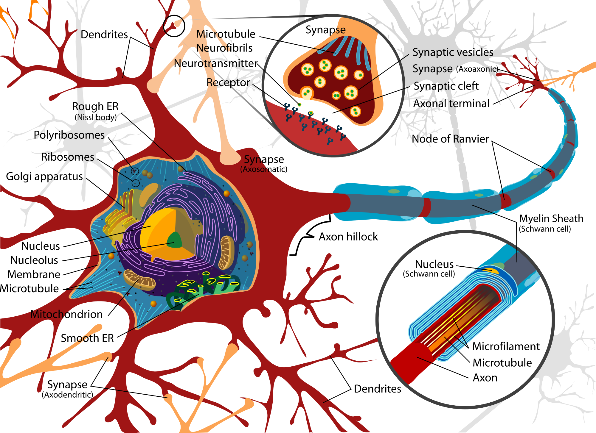
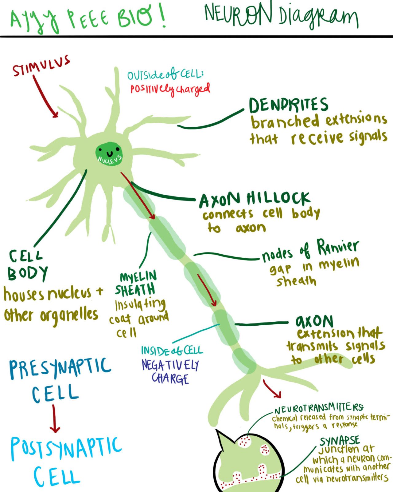
At a synapse, one neuron sends a message to a target neuron—another cell. Most synapses are chemical; these synapses communicate using chemical messengers. Other synapses are electrical; in these synapses, ions flow directly between cells. At a chemical synapse, an action potential triggers the presynaptic neuron to release neurotransmitters.
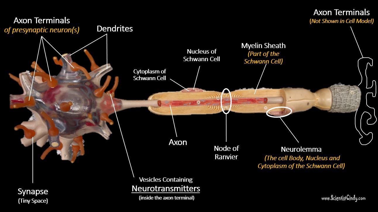
Nervous Tissue SCIENTIST CINDY
Neuron Anatomy. Nerve Cell: Dendrites receive messages from other neurons. The message then moves through the axon to the other end of the neuron, then to the tips of the axon and then into the space between neurons. From there the message can move to the next neuron. Neurons pass messages to each other using a special type of electrical signal.
:max_bytes(150000):strip_icc()/neuron-anatomy-58530ffe3df78ce2c34a7350.jpg)
Neuron Anatomy, Nerve Impulses, and Classifications
The parts of the neuron have been labeled. Your challenge is to write the correct name for each part and explain what it does. If you need some help, visit the web article listed below. Neuron Anatomy Activity. Synapses: Send electrical impulses to neighboring neurons. Myelin sheaths: Cover the axon and work like insulation to help keep.
neuron
3. How to Draw a Neuron Diagram To learn about the structure of the neurons, the students can use a neuron labeled diagram. The students may follow these steps to make their neuron diagram, but the process is complex: 3.1 How to Draw a Neuron Diagram from Sketch Step 1: First, the students need to draw a circle. Based on it, they need to draw a.
Histology of the Nervous System (The Neuron) Part 1
1. Identify the cell type in the above figure Liver Cell Cardiac Cell Nerve cell Skin cell. 2. In the figure, labeled '1' receives impulses from adjacent neuron. It is called the Dendron Dendrite Axon Axonite. 3. In the figure, labeled '2' is the short filaments from the cell body that carries impulses from dendrites to the cell body which is the Schwann cell Axonite Axon Dendron

Neuron Diagram Straight from a Scientist
The cell body of a neuron, also known as the soma, is typically located at the center of the dendritic tree in multipolar neurons.It is spherical or polygonal in shape and relatively small, making up one-tenth of the total cell volume.. The functionality of the neuron is highly dependent on its cell body as it houses the nucleus, which contains the genetic material (DNA) of the cell as well as.
neuron model unlabeled Clip Art Library
Axon. Definition. long, slender projection of a nerve cell, or neuron, that typically conducts electrical impulses away from the neuron's cell body. Location. Term. Cell Body aka Soma. Definition. is the spherical part of the neuron that contains the nucleus. The cell body connects to the dendrites, which bring information to the neuron, and.
